Reasearch Topics: [Video Capsule Endoscopy Analysis]
Computer Vision in Agricultural Engineering and Biodiversity |
Vision-based system
|
Human Computer Interaction |
Human in a Surveillance Camera Network |
| Adaptive Display Control to reduce diagnostic time | |
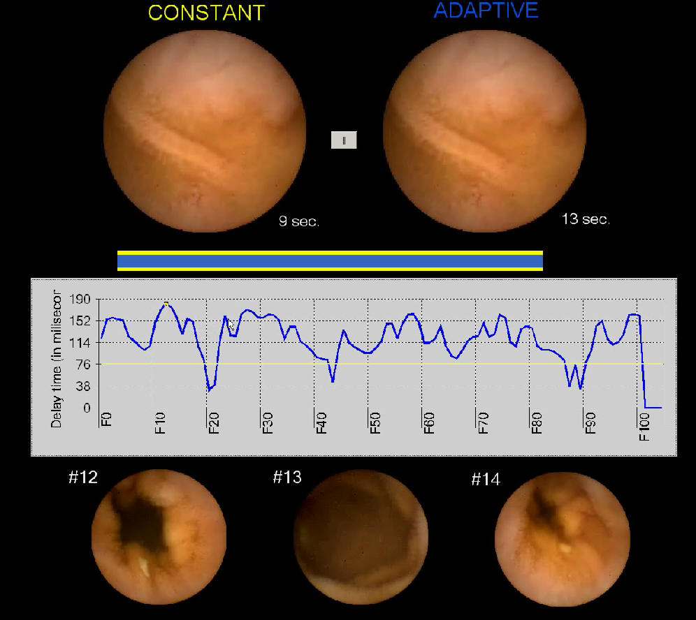 |
Interpretations by physicians
of capsule endoscopy image sequences captured over periods of 7-8
hours usually require 45 to 120 minutes of extreme concentration.
This paper describes a novel method to reduce diagnostic time by
automatically controlling the display frame rate. Unlike existing
techniques, this method displays original images with no skipping of
frames. The sequence can be played at a high frame rate in stable
regions to save time. Then, in regions with rough changes, the speed
is decreased to more conveniently ascertain suspicious findings. To
realize such a system, cue information about the disparity of
consecutive frames, including color similarity and motion
displacements is extracted. A decision tree utilizes these features
to classify the states of the image acquisitions. For each classified state, the delay time between frames is calculated by parametric functions. A scheme selecting the optimal parameters set determined from assessments by physicians is deployed. Experiments involved clinical evaluations to investigate the effectiveness of this method compared to a standard-view using an existing system. Results from logged action based analysis show that compared with an existing system the proposed method reduced diagnostic time to around 32.5 +/- 7 minutes per full sequence while the number of abnormalities found was similar. As well, physicians needed less effort because of the systems efficient operability. The results of the evaluations should convince physicians that they can safely use this method and obtain reduced diagnostic times. |
|
|
| Intestinal Contraction Detection | |
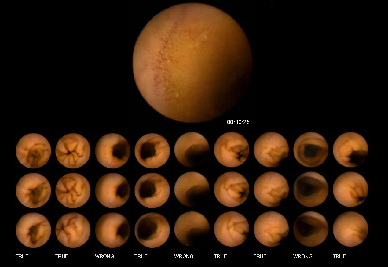 |
Recognizing intestinal contractions from Wireless Capsule Endoscopy (WCE) image sequences provides a non-invasive method of measurement, and suggests a solution to the problems of traditional techniques for assessing intestinal motility. Based on the characteristics of contractile patterns and information on their frequencies, the contractions can be investigated using essential images features extracted from WCE videos. In this study, we proposed a coherent three-stage procedure using temporal and spatial features. The possible contractions are recognized by changes in the edge structure of the intestinal folds in Stage 1 and evaluating similarities features in consecutive frames in Stage 2. In order to take account of the properties of contraction frequency, we consider that the possible contractions are located within windows including consecutive frames. The size of these contraction windows is adjusted according to the passage of the WCE. These procedures aim to exclude as many non-contractions as possible. True contractions are determined through spatial analysis of directional information in Stage 3. Using the proposed method, 81% of true contractions are detected with a 37% false alarm rate for evaluations in the experiments. The overall performance of this method is better than that of previous methods, in terms of both the quality and quantity indices. The results suggest feasible data for further clinical applications. |
|
|
| Segmenting Digestive Organs in Video Capsule Endoscopy | |
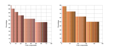 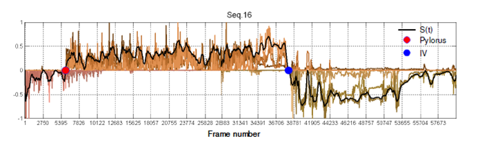 |
This paper presents an efficient method for automatiically segmenting the digestive organs in a Video Cap sule Endoscopy (VCE) sequence. The method is based on unique characteristics of color tones of the digestive organs. We first introduce a color model of the gastrointestinal (GI) tract containing the color components of GI wall and non-wall regions. Based on the wall regions extracted from images, the distribution along the time dimension for each color component is exploited to learn the dominant colors that are candidates for discriminating digestive organs. The strongest candidates are then combined to construct a representative signal to detect the boundary of two adjacent regions. The results of experiments are comparable with previous works, but computation cost is more efficient. |
|
|
| Image-Enhanced Capsule Endoscopy Preserving the Original Color Tones | |
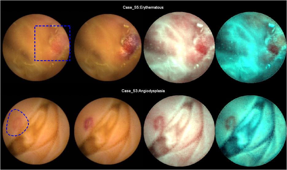 |
This paper describes a technique to enhance a region-of-interest (ROI) in a capsule endoscopy (CE) image. Our aim is to improve distinguishing suspicious regions from normal ones without over enhancing the image. Given a ROI by physicians, the proposed technique enhances the ROI so that the enhanced region remains as natural as possible. To achieve this, we utilize image features relating to a specific color space dedicated to gastrointestinal (GI) wall regions. The proposed approach starts by utilizing a self-organizing map to handle GI wall color components. The goal of preserving the original color tones is obtainable by using a histogram equalization technique in the proposed color space. As a result, the enhanced images only contain color components inferred from the color gamut of the human small bowel. The results of the proposed method are judged in terms of visual appearance by comparison with images obtained by the conventional enhancement technique. |
|
|
| Abnormal regions detection and tracking | |
|
|
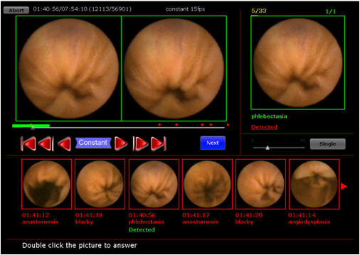
The precise tracking of an abnormality in the gastrointestinal tract is useful for medical training purposes. However, the gastrointestinal wall deforms continuously in an unpredictable manner, while abnormalities lack distinctive features, making them difficult to track over continuous frames. To address this problem, we propose a tracking method for capsule endoscopy using the surrounding features of abnormalities. By applying triangular constraints using an affine transformation, we are able to track abnormalities that do not have distinctive features over consecutive image frames. We demonstrate the efficacy of our approach using eight common types of gastrointestinal abnormalities. |
|
|
| Education System for Reading Video Capsule Endoscopy | |
 |
The interpretive skills of medical doctors and medical technologists who examine video capsule endoscopy (VCE) in clinical practice are usually improved through hands-on courses. Such courses require that a large volume of cases be undertaken as part of the training, and they thus consume a considerable amount of the trainees’ time. This paper describes an e-learning system that reduces the training time in addition to enhancing the quality of the educational process with regard to reading VCE. To achieve this goal, we focused on organizing training courses in order to appropriate for the laborious conditions that exist when reading VCE. The designed courses help the trainees acquire knowledge of abnormal regions and become familiar with reading VCE before taking examinations under conditions similar to those actual clinical practice. The proposed training modality was developed as an e-learning application on the World Wide Web. Thus, it can be easily extended to a wide range of trainees. In the experiments, 20 participants completed the self-learning training procedures in approximately 3 hours. The proposed system is much faster than conventional handson courses, which require a minimum of 8 hours. Furthermore, the trainees’ learning performances in the final examinations confirmed that the proposed system is particularly effective for inexperienced examining doctors. |
|
|